


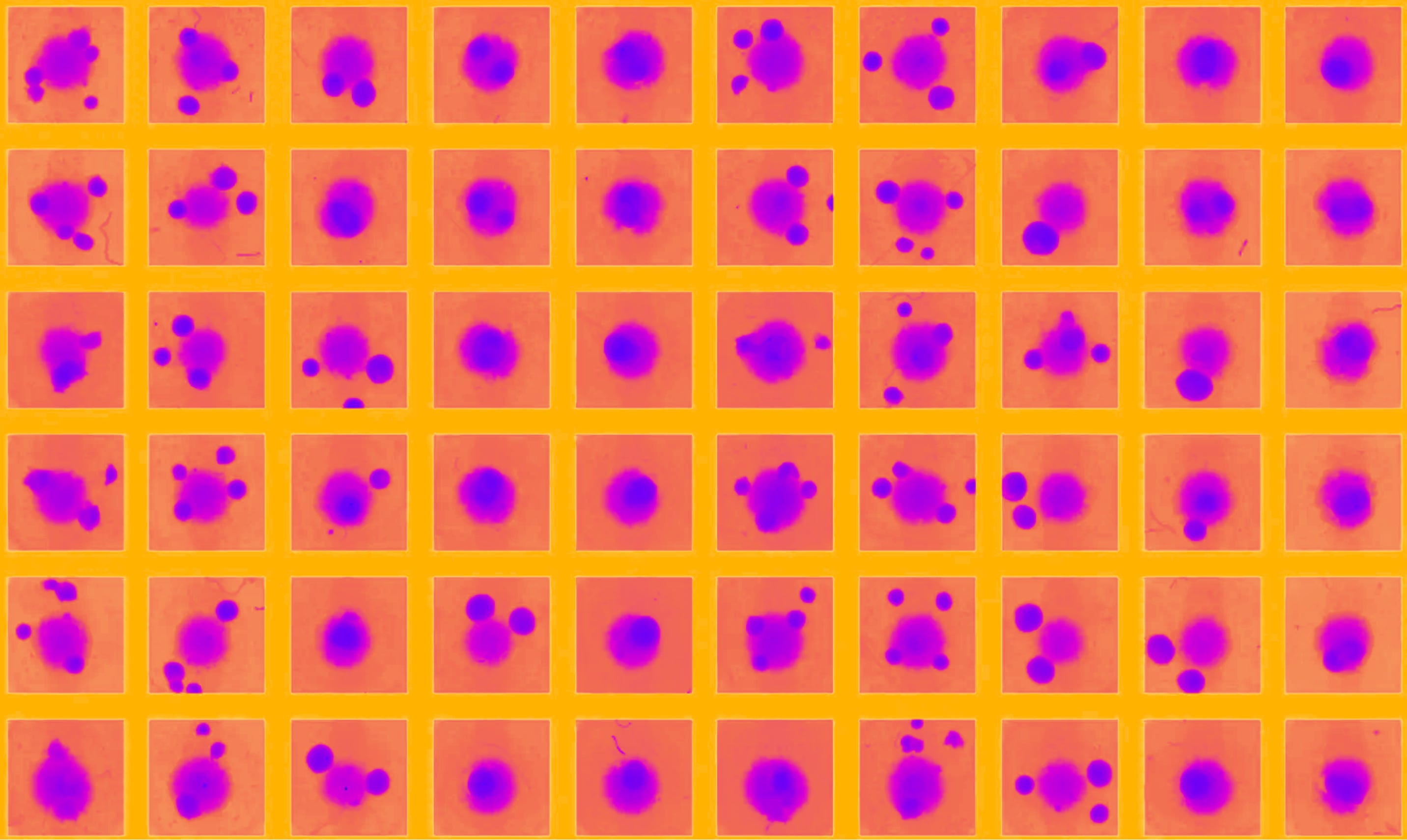
Living organisms are the most complex systems produced by nature. The logic of their inner workings remains one of the great, enduring mysteries of science. Over the past century, the study of biology has produced extraordinary strides, from the discovery of DNA, to gene expression, and the development of increasingly sophisticated theories of disease, enabling us to radically expand our understanding of life. Yet even so, fundamental questions, even at the basic cellular level, continue to stump us. We still lack mechanistic understandings of how cells reliably metabolize, divide and specialize. For all intents and purposes, even these smallest of biological systems remain something like black boxes.
Of course, modern tools have given us unprecedented visibility into this microscopic world. Microscopy itself, transcriptomics, metabolomics, and proteomics let us observe mRNA dynamics, metabolic changes, protein expression, respectively, in great detail. But observation has not yet led to comprehension. The logic and interplay of these components, for the most part, eludes us and the mystery is only exacerbated when scaled to the level of tissues, where millions of interrelated interactions produce complicated, emergent phenomena like coordinated function and differentiation. At the level of the organism, the complexity reaches its apex, and we are left with a system that, simply put, resists direct explanation.
This state of affairs has made the field of medicine, in particular, a uniquely difficult scientific discipline. Nearly all biomedical investigation is conducted with something like a blindfold on. We have been forced to draw conclusions and propose interventions for systems we cannot fully describe. And to complicate the matter further, every organism is unique, which means phenomena like diseases rarely manifest in precisely the same way across different individuals. This is a significant wrench in the scientific method itself, which demands reproducibility.
Until recently, the best solution we had was to anchor on control and standardization. In the face of so many moving parts and variables in flux, the history of biomedical research has narrowed on creating standardized, repeatable model environments where experiments could be performed while minimizing as much noise as possible from confounding variables. And for more than a hundred years there has been one such model that became king in the medical world — the mouse.
Between 30 million to, by some estimates, 100 million rodents are used in biomedical research across the United States and Europe every year. These animals are not creatures captured in the wild and stuffed into cages for medical experimentation, but rather extremely specialized genetic strains, bred selectively over decades and modified to be genetically identical to each other. Mice, as organisms, have some nice properties for mass study. Their gestation cycles are short, they can produce enormous litters, and through careful artificial breeding, the amount of genetic drift exhibited from generation to generation can be suppressed greatly. Their lives are led in sterile, controlled conditions, where exposure to environmental factors that most regular organisms would encounter throughout their lives, like pathogens or dietary variation, are kept to an absolute minimum. It was this industrial mass production of genetically identical organisms that, for a long time, made controlled medical experimentation possible at all.
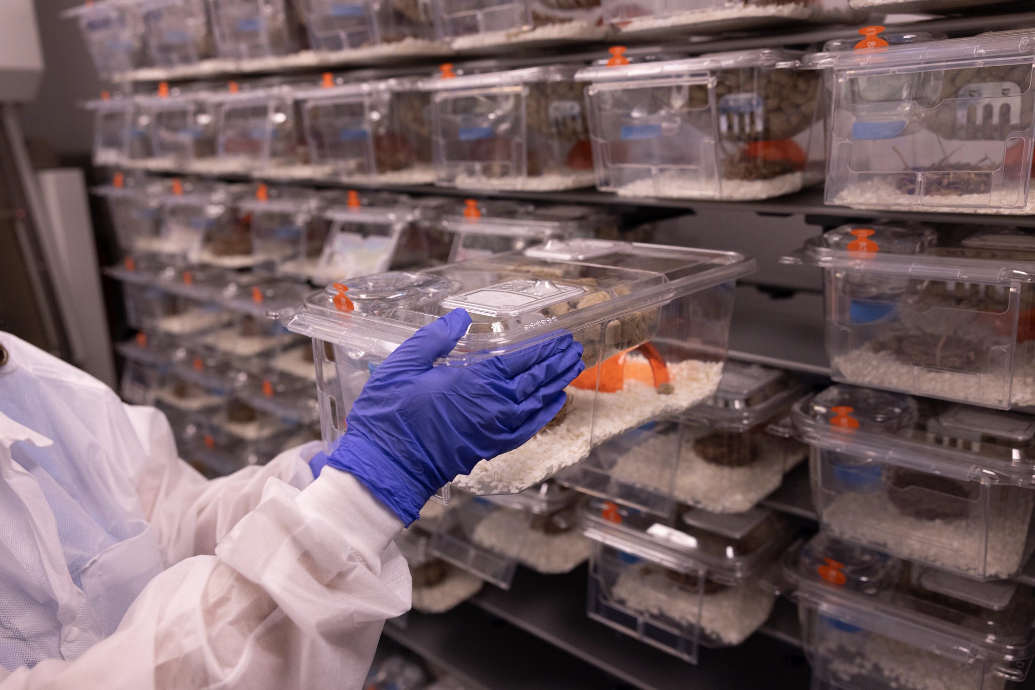
For the better part of a century, nearly every drug that has come to market began with an experiment on mice. But for every drug that has achieved commercial success, thousands of others have failed. For all the reasons that make genetically engineered mice nice vehicles for controlled experimentation, there are countless others that have doomed this path to a scientific dead end.
Mice simply do not get most of the diseases we are interested in studying in humans. They do not get Alzheimer’s, nor Parkinson’s, nor depression. And yet so much of academic experimentation focused on these diseases is conducted in the mouse model. These diseases are induced artificially through genetic knockouts or forced inflammatory states until the animal exhibits symptoms that vaguely resemble the human condition. Is it any surprise, then, that 95% of clinical trials fail after leaving the animal stage, vaporizing billions of dollars of investment per year?
Even among the drugs that do survive this gauntlet and get to market, the efficacy is uneven. A treatment that works well for one patient may fail entirely in another with the same diagnosis. Side effects proliferate all because the precise molecular operation remains only partially understood, incapable of predicting how diverse patient populations will respond even after clinical trials.
This realization, that we have been optimizing for the wrong substrate, has sparked a quiet revolution in how we think about drug discovery. Over the past decade, billions of dollars have flooded into efforts to build better biological models. Some researchers have focused on designing better molecules computationally, using advances in protein structure prediction to engineer drugs that bind more precisely to their targets. Others have turned to high-throughput cell screening, training machine learning models on vast libraries of cellular perturbations. These are impressive technical achievements, and they have advanced our understanding of biology considerably.
Still, a fundamental gap remains. A molecule that binds perfectly to its intended target can still fail in patients, whether due to off-target effects, unexpected immune reactions, or simply because the disease context is more complex than any simplified model can capture. A model trained on immortalized cell lines — cells that have been grown in laboratories for decades, far removed from their original tissue context — may predict those cells well, but tell us little about how a drug will behave in a living patient. There is a big difference between describing what biology looks like and predicting what a drug will do inside a human body. Most of the field’s recent progress, impressive as it is, has been in description.
The limitation, clearly, is in the data itself. The field has grown increasingly sophisticated at building models, but the underlying training data often comes from systems that bear only a passing resemblance to human disease. This is not a criticism of any particular approach. It’s simply a recognition that the most pressing question in drug development is not “can we design a better molecule?” but “will this drug work in this patient?”
A Parallel Approach
This is a bewildering status quo, and it was particularly frustrating to Robert DiFazio. As a graduate student studying tuberculosis, he was astounded by the field’s overwhelming reliance on mouse models. Mice do not get tuberculosis, and when the infection is forced, the resulting disease follows a totally opposite pathological course than is seen in humans.
In any case, Robert wanted to understand immunity at the level of populations. Even if you learned something about how tuberculosis operates in mice, a single inbred strain would never tell you how a disease behaves across thousands of different immune systems. After his PhD, he joined an initiative at Stanford building experimental models out of human tissue, where he came across early work on immune organoids.
Organoids had emerged only a decade prior. In 2009, Hans Clevers and colleagues in the Netherlands showed that adult stem cells isolated from the intestine, when embedded in a supportive gel and supplied with the right environmental cues, could self-organize into miniature gut-like structures.
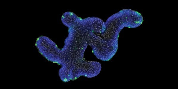
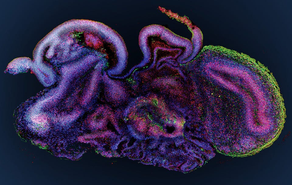
Until then, most studies of cell biology stripped tissues of their native context. Cells were grown on two-dimensional plastic dishes, divorced from the spatial organization that gives organs their form and function. Clevers’ work showed that, under permissive conditions, cells could reconstruct much of that architecture on their own. Similar efforts soon followed. Researchers grew retinal organoids capable of sensing light, and liver buds that metabolized drugs, and even brain organoids, which formed layers mimicking the brain in early development.
Many of the tissues scientists most wanted to study, especially the brain, were impossible to sample from healthy patients. Here, induced pluripotent stem cells offered a way around that constraint. A skin or blood cell could be rewound into an undifferentiated state, then coaxed forward again into almost any tissue. So in principle, one could grow a tiny human brain in a dish, carrying a patient’s exact genetics. In practice, doing so proved challenging.
No one fully understood how exactly cells organized themselves into these structures, and attempts to manually steer their differentiation yielded inconsistent results. Early organoids, especially those derived from induced pluripotent stem cells, varied wildly from batch to batch. If you tried to generate a forebrain organoid from one donor's cells and then applied the exact same protocol to cells from a second donor, you would often end up with a completely different region of the brain. The lack of reproducibility made these unusable as scientific models. Academics sidestepped the issue by cherry-picking the few cell lines that behaved predictably, but that only reinforced industry skepticism that organoids were inherently unreliable.
At the same time, Juliana Hilliard was working at the Knoblich Lab in Austria, studying brain organoids and attempting to tackle this very problem. She wanted to understand how cells could be guided down reproducible pathways so organoids from different donors would behave the same way. Without that, the entire field would be stuck.
Juliana, by her own admission, never liked the messiness of biology. “Biology never gave you a right answer,” she says, “which is tricky.” Instead, she gravitated toward engineering, where systems could be constrained and made to work. The possibility that living tissues might one day be engineered with the same rigor as machines was exactly what drew her to organoids.
Robert and Juliana were moving through adjacent intellectual terrain, dissatisfied with incrementalism, and circling the same unanswered question — how do you stop approximating human biology and build experimental systems that are human?
When the pair finally met in San Francisco, they realized the field’s problems all traced back to brute-forcing organoid assembly with induced pluripotent stem cells. If the goal was to create an experimental platform to reproducibly understand disease across patient populations, why not start directly from primary human tissue, which already carried the instructions for self-assembly, and focus on the immune system, which itself encodes a patient’s entire history, from genetic predispositions to idiosyncrasies from past infections?
If it worked, they would have organoids that were consistent within a single donor and comparable between them. These were the insights around which their company, Parallel Bio, was founded.
A New Paradigm for Biology
Medicine succeeds or fails at the level of populations. A drug might help 90% of patients or only 30%, or despite being generally effective, it might also be toxic in 5% of people.
To model this reality, Parallel produces organoids at scale. It selects patient cohorts from a biobank of more than a hundred and eighty donors, matched by age, sex, genetics, prior exposures, and disease state, and builds immune organoids from those exact donors. Drugs are introduced directly into these systems, then read out every which way, from microscopy to single-cell sequencing, cytokine profiling, receptor-level analysis, and functional immune assays. Every result is tied back to the donor’s metadata, so a drug's effect can be read for a single patient or a whole population at once. It’s something no existing model, much less animal model, can do.
Robert and Juliana soon realized the platform they had on their hands was really something that resembled a clinical trial, conducted entirely in a petri dish.
To build it, they turned to biobanks holding vast reserves of patient-derived immune cells and began assembling their first immune organoids, essentially miniature lymph nodes. The results were immediately striking. These lymph organoids contained hundreds of distinct cell subtypes, many of which would have been impossible to engineer by hand, some of which scientists don’t even routinely characterize.
More remarkably, the organoids behaved like real immune tissue. They showed transcriptional responses by day three, produced antibodies by day five, and exhibited the same 21-day antibody peak observed in human vaccination.
When tested retrospectively on TGN1412, a drug that passed animal studies in 2006 but caused near-fatal reactions in six healthy participants during the first hours of a Phase 1 trial, Parallel’s organoids correctly identified the dangerous immune response that animal models had missed entirely. The platform predicted, in a dish, what took a catastrophic human trial to reveal.
The commercial relevance was immediately apparent. Roughly half of all drugs in development target the immune system, and with Parallel’s platform, researchers wouldn’t have to spend five to ten years building and validating disease models. They could simply run experiments directly on human tissue in weeks rather than decades.
Theirs was a technology that could accurately predict drug safety and success before ever entering humans. This meant everything from the use of animal models and eventually even Phase I trials in humans could be bypassed entirely. Gone would be the days of years of effort, and billions of dollars of funding required to sustain incremental progress.
Though traditional academia and industry dragged their feet on acknowledging the paradigm shift under way, public officials quickly took notice. Parallel Bio started in 2021. A year later, Congress passed the FDA Modernization Act of 2022. Until then, results from animal models were needed to open any new Investigational New Drug application, or IND, which required repeated-dose toxicology seen across two mammalian species, usually a rodent and non-rodent model. Non-rodent animal models, typically mini-pigs or non-human primates, however, were trickier to work with. First of all, their genetic makeup was far more variable than mice, their gestation cycles were longer, and they made results far more difficult to standardize at scale. The FDA Modernization Act stated that from then on, animal testing would no longer be a mandatory requirement for IND applications. Instead, sufficiently detailed testing performed through in silico or in vitro approaches, like organoid testing, would be sufficient on their own. In April of 2025, the FDA doubled down on this position, publicly laying out a roadmap to reduce animal testing in preclinical safety studies, and by December, put out guidance aimed at reducing, if not eliminating, non-human primate toxicity studies for certain applications.
Shortly after, pharma started taking note. Parallel’s first major opportunity came in vaccines. They partnered with Centivax, which was working on designing what could become the world’s first universal flu vaccine. Vaccine development is notoriously fraught, with many candidates failing in humans despite promising animal data. Using Parallel’s platform, immune organoids derived from adult donors were exposed to Centivax’s lead program, Centi-Flu. The organoids mounted broad immune responses capable of recognizing multiple influenza strains, including ones not directly represented in the vaccine. For Centivax, this was a breakthrough because it was evidence of human efficacy before a single patient had ever been dosed.
Since then, Parallel has expanded to partnerships with numerous pharmaceutical companies, including three in the Fortune 500, with more than eighty active drug programs running across its platform.
The New Economics of Medicine
Parallel came to see that many diseases we label by organ are, in fact, driven by immune dysfunction upstream. Inflammatory Bowel Disease manifests in the gut, but its causal signals appear in lymph nodes. Rheumatoid Arthritis attacks the synovium, but its impact on the immune system can be modeled without rebuilding the joint. Multiple Sclerosis damages myelin, but the misfiring begins in immune circuitry long before neurons are lost. As more of this immunological iceberg comes into view, it’s clear that the immune system touches nearly every domain of disease, from heart disease, neurodegeneration, and even aging itself, which emerging evidence suggests may actually be heavily immune-regulated.
If this is true, then whoever can model the human immune system at scale, on real patient tissue rather than simplified cell lines, and across the full diversity of human biology, will understand disease in a way no one has before. This is the vision that now animates Parallel Bio.
It is difficult to overstate the implications of this shift. Today, pharmaceutical development typically takes close to a decade and costs on the order of two billion dollars for each drug that reaches the market, an outcome achieved by only 5% of candidates that enter clinical trials. The overwhelming majority fail only after years of work, vast capital expense, and exposure of patients to compounds that had very little chance of ever succeeding.
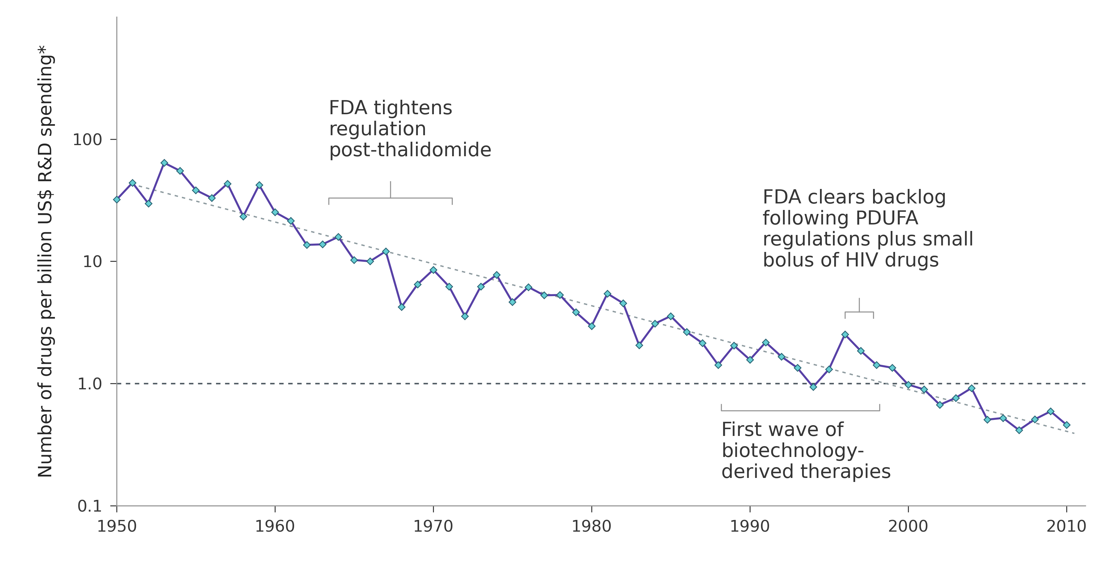
Now imagine a world in which that attrition happens earlier, before human trials ever begin. Imagine a pipeline where weak candidates are filtered out at the level of human biology, rather than through expensive and ethically fraught experimentation in people. In such a system, the odds could be reversed. The small fraction of drugs that reach clinical trials would do so because they were already known to work in human tissue, not because they had merely passed through a long procession of imperfect proxies.
Robert compares the potential of this shift to what SpaceX did for launch economics, namely driving the cost of putting mass into orbit from more than $20,000 per kilogram to just a few hundred. Drug development, long constrained by cost, risk, and time, could undergo a similar compression. Timelines would shrink. Capital efficiency would improve. And downstream, patients would see not only faster access to therapies, but drugs that are safer, more effective, and designed with human biology in mind from the start.
And perhaps even this doesn’t quite capture the full picture of what’s possible. After all, one of the most consequential tools reshaping how we study biology itself is artificial intelligence. For the first time, we have a method capable of absorbing the full complexity of a biological system and reasoning over it directly, rather than forcing us to isolate individual components and extrapolate outward.
But models are only as good as what they’re trained on, and so far that has mostly been systems far removed from human disease, like immortalized cell lines, healthy cells, or flat cultures with none of the architecture of real tissue. Models trained this way can predict these systems well, but real patients poorly. What’s been missing all this time is data from the place disease actually happens, in tissues, in organs, and in the way they interact over time.
Parallel is building that dataset now. Their conviction is that with enough human-grounded data, it becomes possible to build foundational models of biology that invert the logic of biomedical inquiry. Instead of starting with narrow hypotheses and testing them in simplified systems, we can start with observations that preserve the complexity of biological function and let structure, mechanism, and insight emerge from there. Every disease model strengthens the foundation. Every donor expands its reach. Every response to a perturbation adds signal. And clinical validation, the one thing that cannot be replicated in silico, then closes the loop. In doing so, Parallel is making it possible to hold biological complexity in our hands, and finally understand life at a depth that has long been out of reach.